Modality Requirements at a Glance
Three distinct modalities dominate clinical nuclear imaging, each with a unique scintillator specification profile.
Positron Emission Tomography (PET)
PET detects coincident 511 keV annihilation photons. Speed is the primary constraint. A short decay time reduces random coincidences and allows tighter timing windows, which directly improves image signal-to-noise. High density and high atomic number (Z) are equally critical: they stop 511 keV gammas efficiently in thin detector elements, keeping scanner diameter manageable and sensitivity high. Light yield matters, but decay time and stopping power take priority over raw photon count.
Single-Photon Emission Computed Tomography (SPECT)
SPECT uses collimation rather than coincidence, so it does not impose the same sub-nanosecond timing requirement. Energy resolution becomes more important here, because the system must discriminate the primary photopeak from scattered photons. A modest decay time (hundreds of nanoseconds to a few microseconds) is acceptable. Good energy resolution and high light yield allow finer energy windowing, reducing scatter contamination and improving lesion contrast.
Computed Tomography (CT)
CT operates in current-integration mode with X-ray sources, not radioactive tracers. The demands shift: very low afterglow (no phosphorescence after the X-ray pulse ends), high density for X-ray stopping power, and compatibility with photodiode readout. A scintillator with slow primary decay is acceptable provided afterglow is negligible. Hygroscopicity is a concern in production arrays, so non-hygroscopic materials are strongly preferred.
ScintIQ Materials for Medical Imaging
Four ScintIQ materials with published datasheets serve medical imaging, and a fifth, CdWO₄, is available on request. Their key properties are summarized below; full specifications appear on each material's data sheet.
| Material | Density (g/cm³) | Decay Time | Emission (nm) | Light Yield (NaI = 100 unless stated) | Hygroscopic | Primary Imaging Use |
|---|---|---|---|---|---|---|
| LYSO(Ce) | 7.2 | 50 ns | 420 | 76 | No | PET (dominant choice) |
| BGO | 7.13 | 0.3 μs | 480 | 15–20 | No | PET (legacy / cost-sensitive) |
| GAGG(Ce) | 6.6 | <90 ns | 520 | ~50,000 ph/MeV | No | PET, SPECT, gamma camera arrays |
| CsI(Tl) | 4.51 | 0.6 / 3.4 μs | 550 | 45 | Slightly | SPECT, gamma camera, general |
| CdWO₄ | on request | on request | on request | on request | No | CT arrays, high-flux X-ray |
Note: Relative light yield is photoelectron yield versus NaI(Tl) = 100 on a bialkali PMT; the GAGG(Ce) datasheet states absolute light output only. CdWO₄ specifications and sample quantities are available on request.
Understanding the Key Tradeoffs
LYSO(Ce): The PET Standard
LYSO dominates modern PET because it combines the two properties that matter most: density (7.20 g/cm³) and speed (50 ns decay). High density means more 511 keV photon stopping power per unit volume, allowing thin detector elements and compact scanner geometries. The 50 ns decay supports narrow coincidence windows and time-of-flight reconstruction, reducing random coincidences and improving sensitivity. LYSO is non-hygroscopic and compatible with SiPM readout, which has replaced PMTs in production time-of-flight PET. Its relative light yield of 76 and 8 percent energy resolution support the energy windowing PET needs without being the highest available. LYSO is the first material to specify for any new PET detector design.
BGO: Density at Lower Cost
Bismuth germanate (BGO) was the PET workhorse before LYSO became widely available. Its density (7.13 g/cm³) and Z are comparable to LYSO, and it has no afterglow. The limitation is light yield: at 15 to 20 relative to NaI(Tl), BGO produces far fewer photons per gamma event, which limits energy resolution (typically 8 to 12 percent at 662 keV) and timing; SiPM readout is feasible and increasingly used in compact designs. BGO also decays slowly (300 ns), which limits count-rate performance. It remains relevant in budget-conscious configurations or legacy replacement programs where the original scanner was designed around BGO array geometry.
GAGG(Ce): Flexible Across Modalities
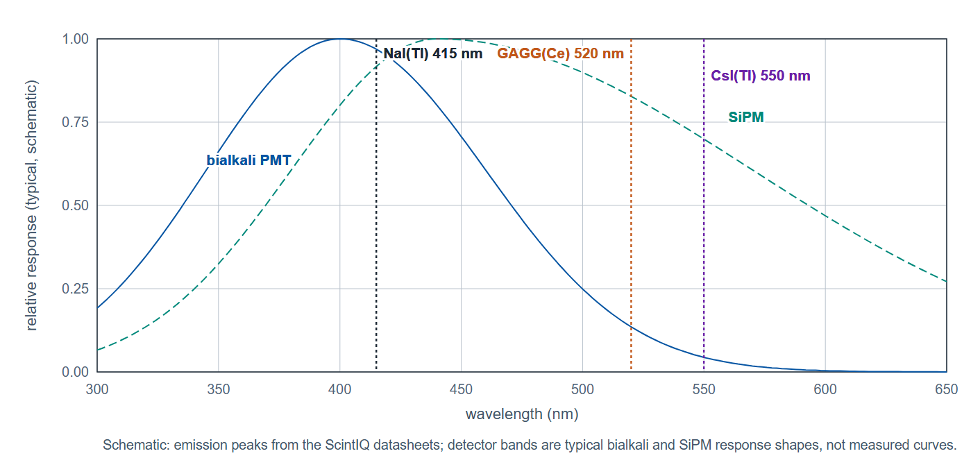
GAGG is a newer oxide scintillator with a useful combination of density (6.6 g/cm³), speed (primary decay under 90 ns), and green emission (520 nm) that suits SiPM readout. It does not match LYSO's stopping power or BGO's legacy installed base, but it fills a practical role in SPECT detector arrays, small-animal PET, and gamma camera applications where SiPM compatibility and radiation hardness are valued. GAGG is also non-hygroscopic, which simplifies array assembly. Its light output of about 50,000 photons per MeV and about 6 percent energy resolution with a high-QE PMT support energy discrimination at SPECT energies (140 keV for Tc-99m).
CsI(Tl): SPECT and Gamma Cameras
NaI(Tl) is the Anger gamma camera scintillator; CsI(Tl) is the photodiode-coupled choice for CT arrays, flat-panel detectors and compact cameras. Its 550 nm yellow-green emission is a direct match to photodiodes and silicon sensors, and it delivers 45 relative light yield with only slight hygroscopicity. The slower decay (600 ns and 3.4 μs) is acceptable where coincidence timing is not required. CsI(Tl) crystals can be grown in large monolithic slabs or segmented arrays, and the crystal is mechanically rugged with no cleavage plane. For compact cameras that use SiPM arrays, CsI(Tl) offers a well-proven path.
CdWO₄: CT Arrays and High-Flux X-Ray
Cadmium tungstate is the non-hygroscopic CT array material, chosen for its density and low afterglow: after each X-ray pulse the scintillator must return to baseline before the next measurement, and afterglow smears slice data. Its slow decay is no penalty in current-integration CT readout, and its emission suits silicon photodiodes. CdWO₄ is not in the published ScintIQ datasheet set; Berkeley Nucleonics quotes it on request in custom bars and arrays.
Readout Options and System Integration
All ScintIQ materials for medical imaging are available in a range of crystal sizes, surface finishes, and housing configurations. The choice of readout device interacts directly with emission wavelength and light yield.
- SiPM readout: Preferred for LYSO and GAGG in modern PET and compact SPECT. SiPMs operate at low bias voltage, are MRI-compatible, and scale well to pixelated array formats. The ScintIQ SiPM Readout module pairs with LYSO and GAGG crystals for integrated detector assemblies.
- PMT readout: Remains standard for large-area SPECT detectors and gamma cameras using CsI(Tl) or NaI(Tl). Position-sensitive PMTs provide good spatial resolution across large crystal areas. ScintIQ assemblies include crystal-to-PMT optical coupling with light guide options.
- Photodiode readout: The natural choice for CsI(Tl) and CdWO₄ in CT, where the 550 nm emission matches silicon photodiode quantum efficiency. Photodiodes operate at low voltage without high-voltage supplies, simplifying array electronics.
Detector housing, reflector materials, optical coupling grease, and hermetic encapsulation are all configurable through the ScintIQ custom detector program. Crystal dimensions, pixel pitch, and array formats are specified per project. Contact Berkeley Nucleonics engineering to discuss your detector geometry and count-rate requirements.
ScintIQ Material Data Sheets
Full specifications, energy resolution data, and available configurations for each material are documented in the ScintIQ series data sheets:
CdWO₄ specifications and sample quantities are available on request. Use the ScintIQ material comparison tool to filter the ScintIQ materials by density, speed and application.
Medical imaging applications often require custom crystal dimensions, array formats, or specific reflector and encapsulation configurations. Our team can provide application-specific recommendations, prototype crystals, and pricing.
Email: info@berkeleynucleonics.com · Phone: 800-234-7858