Inconsistencies in measuring dose rate and specifying accuracy can become a frustrating experience. This paper will uncover some of the discrepancies in making these measurements as well as misleading assumptions when using different instrumentation. Dose rate measurements can be performed for different types of radiation (e.g., alpha, beta, gamma and X-rays). This paper will confine its discussion to gamma and X-ray (photon) radiation since we will relate these measurements to the use of scintillation and solid-state detector spectroscopy.

Defining the Terms used for Dose Rate
There are two terms frequently used in defining dose rate: Exposure rate and Equivalent rate [1]. Exposure rate is defined as the amount of ionizing radiation per hour in a person’s vicinity, measured in Roentgens per hour (R/h) [2]. Equivalent rate refers to the biological effect (absorbed dose rate in the human body) from exposure to that radiation, measured in Sieverts per hour (Sv/h). The Sv/h is the international standard (SI) which is quantified as: 1 Sv/h equals 100 roentgen equivalent man per hour (100 rem/h). Represented as:
1 Sv/h = 100 rem/h or 100 nSv/h = 10 µrem/h
Where 100 nSv/h is often a typical background measurement.
The concept of dose equivalent (H) was introduced to more adequately quantify the biological effect of all types of radiation exposure. The standard dose equivalent is usually represented by H(10) which indicates that this biological dose is absorbed in the body to a depth of 10 mm under the skin. It should be mentioned that rem is still very popular in the United States but the Sievert is growing in popularity especially within scientific and government agencies. For example, the EPA in the United States has used the SI as, nSv/h, for easy comparison with all international monitoring stations.

Techniques and Considerations for Accurate Measurements
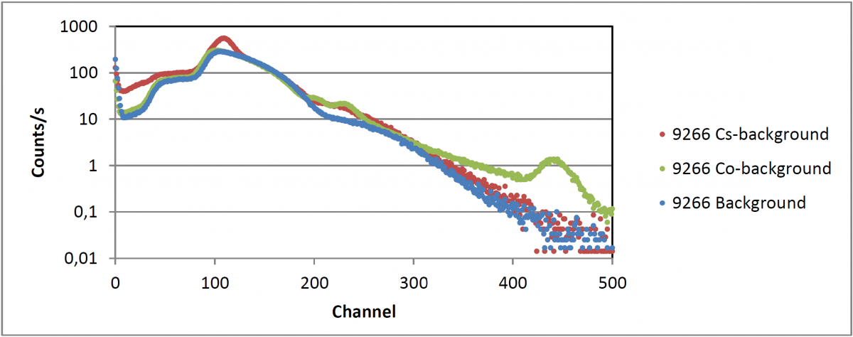
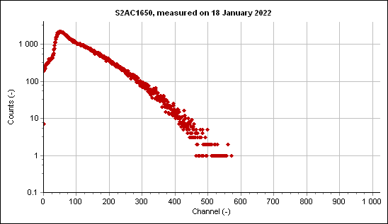
Scintillation detector instrumentation must be calibrated for gamma efficiency related to photon energy in the spectrum. The efficiency curve is then incorporated into the firmware of the instrument. Many factors determine this efficiency curve such as the type of detector, detector size, scintillator type, losses in the detector window and other factors affecting low and high energy (including energies above 1500 keV). Today the miniaturization of sophisticated microprocessors and firmware have allowed outstanding performance of complicated tasks not possible many years ago. This can permit more efficient processing of variables at high count rates. As indicated above, high energy losses can play a part in the accuracy of dose rate measurements. As the photon energy increases the higher energy photons can become a significant factor in determining the total dose rate since this energy is directly proportional to the dose rate. Therefore, the instrument must be sensitive and capable of good linearity across all energies, generally zero to 3000 keV. Further discussion on this will appear under the subject of ion chamber measurements.
There are also many considerations the user must be aware of when performing measurements. Primarily, the solid-state detector must be in the form of a right circular cylinder. Other detector forms do not allow total photon absorption within a solid angle. Since the radiation from a point source is spherical (4π) the right circular cylinder must also be positioned directly in front of the source (on the centerline with the source) to avoid errors in geometry. When considering the solid angle on the detector, it becomes obvious that sources that begin to depart from a point source will also cause measurement error. Large detectors (e.g., 4x4x16 inches) are chosen for maximizing sensitivity, not for dose rate accuracy. These unconventional detector shapes are generally calibrated so that ambient dose rate readings will represent true background measurements.
When making dose rate measurements and calculating the equivalent dose rate at a measured distance from the detector, there are several factors that need to be considered. For distances that are relatively close to the detector it is important to know the exact location of the detector inside the enclosure (behind the detector window). It is also important to accurately measure the distance to the detector because of the significant effect from the inverse square law [3]. Even though measurements can be made with small sources up close (and even on the detector window), the accuracy of the measurement will suffer. This is because the solid angle will become very large, limiting full absorption of the photons near the circumference of the detector. For this reason, most manufacturers suggest positioning the detector no closer to the source than five-times the diameter of the detector for accurate readings. This is especially true for high energy sources which require adequate detector volume to ensure good photon interaction which will limit Compton scatter and losses in full photopeak response.
There are certain precautions that are important when setting up a good measurement area. The measurement area (including table) should be free of metal which can produce photon scatter, sometimes referred to as shine. When picking a source for calibration use a mono energetic isotope (e.g., Cs-137 with its moderate energy of 662 keV). Be aware that low energy sources (e.g., Am-241) can have appreciable absorption in air when measuring a source at long distances from the detector.
When powering up an instrument, especially for the first time, be sure there has been adequate time for warm up. An instrument that has been stored or operated in cold weather and then brought inside to calibrate must be allowed to reach room temperature. After calibration, remove all sources in the vicinity of the detector before taking a background. It has been observed that users may forget to remove the calibration source (usually Cs-137) before taking a background. When this happens, the instrument will lack sensitivity in the 662 keV region and in addition cause large dose rate errors at that energy.

Ionization Chamber Detectors
Air-filled ionization chambers are one of the oldest methods used to measure gamma-ray exposure rate. Measuring exposure rate with an ionization chamber is well suited for this application since exposure is defined in terms of the amount of ionization charge. These chambers are rather large and bulky; however, the size can be limited to some extent. It is impractical to make a chamber large enough to handle secondary electrons since they can range in air up to several meters. Therefore, a compensation called “free-air” is used [4]. The parallel plate ionization chamber is the most common instrument and is used in many countries. The agencies in some countries (e.g., throughout China) standardize on the ionization chamber as the most accurate and therefore the bench-mark in comparison to all other dose rate instruments. This can become a problem in many regions where radioactive outcroppings have substantial deposits of radium and thorium. This is especially true in Asian countries where there are high levels of actinides at sea level and therefore high levels in building materials. BNC has performed scintillator measurements on building walls, concrete floors and other structures in Korea, Japan and China where the ambient dose rate can easily be three times the level commonly experienced in the U.S. Exposure rates of sources in some concrete buildings can produce measurement errors of 20% or more when performed with ionization chamber readings. This is because radium and thorium have high energy photons well above the linearity specifications for these instruments; especially for thorium background with a significant dose rate from photons at 2615 keV (from Tl-208). It has been noted that the specifications of these ionization chambers show a prominent decrease in sensitivity above ~1700 keV, whereas, a 3×3 inch scintillation detector has much better linearity and photon absorption at these high energies.
Often it becomes difficult for the manufacturer to specify dose rate accuracy when not knowing if the user appreciates all the above considerations. This becomes apparent when we see accuracies specified at +/- 20% or more, knowing it could be better or worse.
References
[1] G. Knoll, Radiation Detection and Measurement, Second Edition, 62. (1989)
[2] Further definitions and extensive discussions on these measurements and units are found in the publications of the International Commission on Radiation Units and Measurements. In particular the ICRU Report #19. (1971)
[3] “Newton’s Inverse Square Law as Applied to Radioactivity”, BNC web-site (Berkeleynucleonics.com).
[4] G. Knoll, Radiation Detection and Measurement, Second Edition, 142. (1989)